Most students can name "support" and stop. The skeletal system actually has five distinct functions, and an exam often tests whether you can list and explain all of them. The skeleton is a structural frame, a shield, a system of levers, a blood-cell factory, and a mineral reservoir — all at once. Once you can match each function to the bone tissue that does it, the system stops looking like a pile of bones and starts looking like an active organ.
The Five Functions, in One Sentence Each
Use this list as the skeleton (sorry) of your notes:
- Support. Bones form the frame that holds the body upright and gives soft tissues a place to attach.
- Protection. Bones shield the most vulnerable organs — the skull protects the brain, the rib cage protects the heart and lungs, the vertebrae protect the spinal cord, the pelvis protects pelvic organs.
- Movement. Bones act as levers; muscles pull on them across joints to produce motion.
- Blood cell formation (hematopoiesis). Red bone marrow inside certain bones produces all red blood cells, all platelets, and most white blood cells.
- Mineral and energy storage. Bones store about 99% of the body's calcium and most of its phosphorus. Yellow marrow stores fat as an energy reserve.
A common exam trap: bones do not "make muscles work" — muscles do that. Bones provide the lever; muscles supply the force.
Support and Protection: The Frame and the Shield
The skeleton's most obvious job is structural. About 206 bones in an adult form a continuous frame that lets the body stand upright against gravity, holds its shape, and gives insertion points for skeletal muscle. Without the skeleton you would not be a column; you would be a heap.
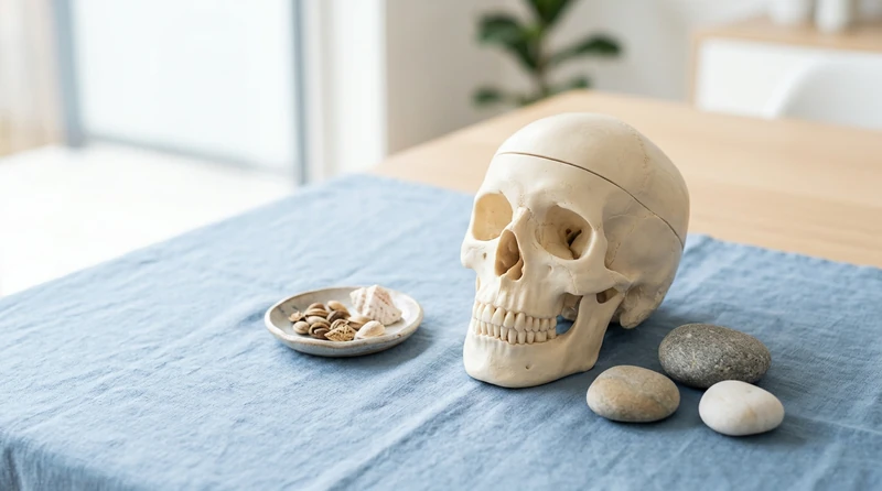
Protection is structural too, but specific. The skull is a closed dome around the brain. The vertebral column is a chain of holes around the spinal cord. The rib cage forms a basket around the heart and lungs, attached to the sternum in front and the thoracic vertebrae behind. The pelvis is a bowl around the bladder, reproductive organs, and lower digestive tract. Each protective structure surrounds the organ it shields almost entirely — the only exceptions, like the eyes, sit in bony sockets that protect them from most angles.
Movement: Bones as Levers
Bones do not contract. They move because skeletal muscles, attached by tendons, pull on them. At every joint, the meeting point of two bones, that pull translates into rotation. Most movements you make are third-class levers: the muscle attaches close to the joint and applies force between the joint (the fulcrum) and the load. A third-class lever sacrifices mechanical advantage for speed and range — your biceps moves your forearm a long distance with a small contraction.
Different joints allow different motions. A hinge joint like the elbow allows movement in one plane. A ball-and-socket joint like the hip allows movement in three planes. A pivot joint like the one between the first two vertebrae lets your head rotate. The bones provide the rigid lever; the joint type determines what the lever can do.
Blood Cell Formation and Storage: The Living Inside of Bone
This is the function most students miss. Inside many bones — especially the ends of long bones, the ribs, the sternum, the vertebrae, and parts of the pelvis — sits red bone marrow, the body's only major site of hematopoiesis (blood cell formation). Red marrow produces red blood cells, platelets, and most white blood cells. In adults, the pelvis, sternum, and vertebrae are the biggest contributors, which is why bone marrow biopsies are taken from those locations.
Yellow bone marrow, found in the shafts of long bones, is mostly fat and serves as an energy reserve. In severe blood loss, yellow marrow can convert back to red marrow to ramp up blood cell production.
Bone is also the body's main mineral reservoir. About 99% of the body's calcium and about 85% of its phosphorus are stored in the matrix of bone. The two are not locked in; they are constantly cycled. Osteoblasts lay down new bone matrix and pull calcium out of the blood; osteoclasts break down bone matrix and release calcium back into the blood. Parathyroid hormone raises blood calcium by activating osteoclasts; calcitonin lowers it by activating osteoblasts. The skeleton is essentially the body's calcium bank, and its balance is critical for nerve and muscle function.
Compact vs. Spongy Bone: Two Tissues, One Bone
Every bone is built from two types of bone tissue, organized differently.
Compact bone (also called cortical bone) forms the dense outer shell of every bone. Its functional unit is the osteon (Haversian system): concentric rings of bone matrix wrapped around a central canal that carries blood vessels and nerves. The structure looks like a stack of microscopic straws aligned along the bone's long axis. Compact bone is strong under compression and bending — it bears most of the body's weight.
Spongy bone (also called cancellous or trabecular bone) sits inside the ends of long bones and inside flat bones. It is a lattice of bony struts called trabeculae, with marrow filling the spaces in between. The struts are arranged along the lines of stress the bone receives — a hip bone is a textbook example, with trabeculae oriented to handle the load of standing and walking. Spongy bone is lighter and absorbs shock, and the spaces between its struts house red marrow.
A long bone like the femur has compact bone in the shaft, spongy bone in the ends, yellow marrow in the central cavity, and red marrow in the spongy spaces. The same two tissues, different arrangements, doing different mechanical and metabolic jobs.
Getting Help
The trickiest part of the skeletal system is usually memorizing all 206 bones — that is the topic of how to memorize the bones. For more anatomy walkthroughs, see the full set of Anatomy & Physiology study guides.
Conclusion
The five functions of the skeletal system are support, protection, movement, blood cell formation, and mineral storage — and each maps onto a specific feature of bone. The hard outer shell of compact bone provides the support, the protective domes shield organs, the joints turn bones into levers, the red marrow inside spongy bone makes blood cells, and the calcium-rich matrix is the body's mineral bank. Together they make bone a living, working organ, not just a frame.